Strain Snapshot
Why Dual IL4/IL4R Humanization?
IL4 signaling through the type I receptor complex (IL4 + IL4Rα + γc) drives Th2 immune responses. Human therapeutics targeting IL4 or IL4R require both human ligand and receptor for proper binding and signaling.
The hIL4/hIL4R model humanizes both IL4 and IL4R to provide:
- Human IL4 ligand binding to human IL4R receptor
- Authentic type I receptor complex formation
- Proper downstream signaling through humanized pathway
- Normal mouse immune system architecture
This dual humanization enables testing of anti IL4/IL4R therapeutics with their native human targets in a fully immunocompetent model.
Validation Data
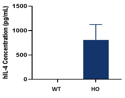
The homozygous KI mice express hIL4 in serum after treatment with concanavalin.
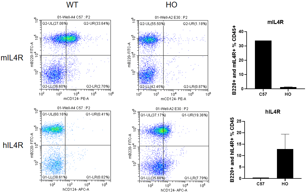
The homozygous KI mice express hIL4R in the spleen, and the WT mice only express mIL4R.
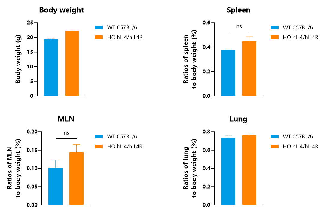
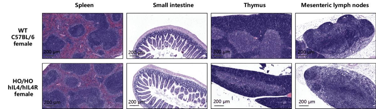
Abbr. HO, homozygous; WT, wild type; MLN, mesenteric lymph nodes.
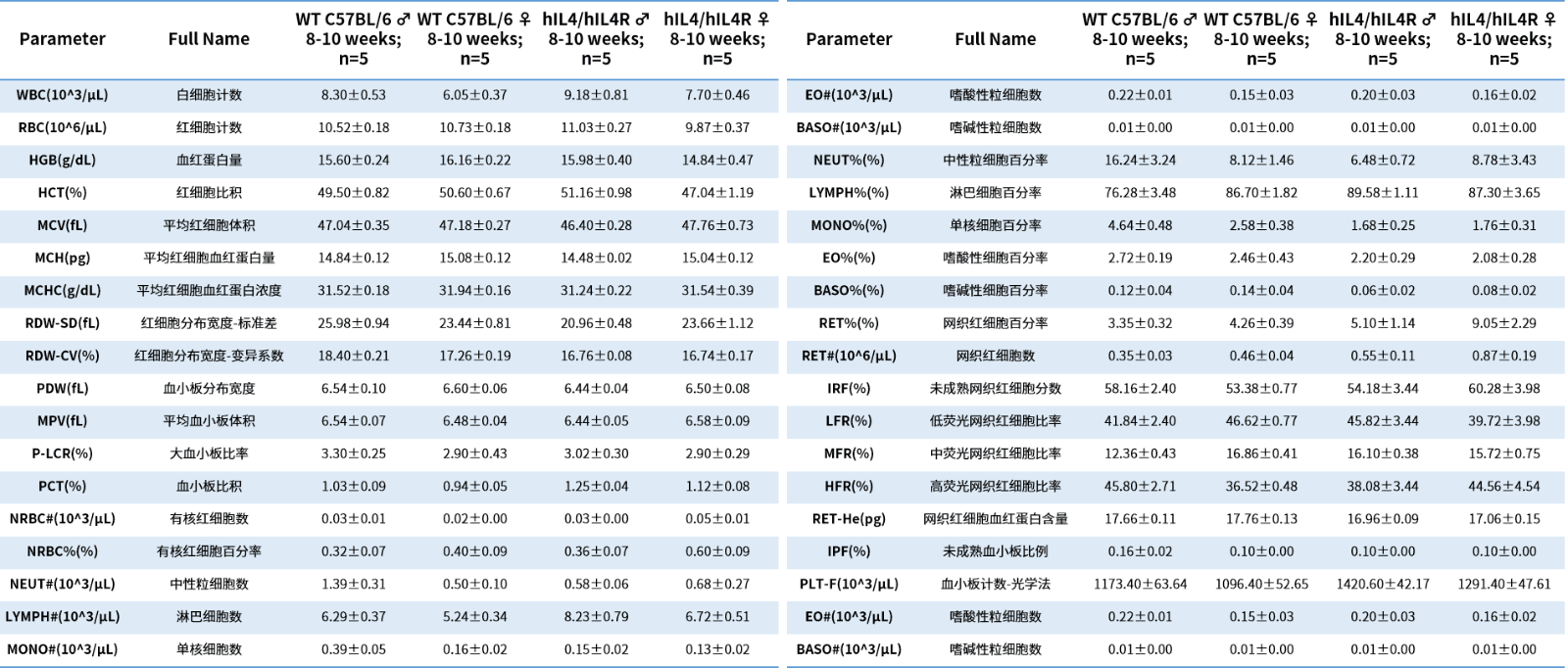
Abbr. WT, wild type; HO, homozygous.

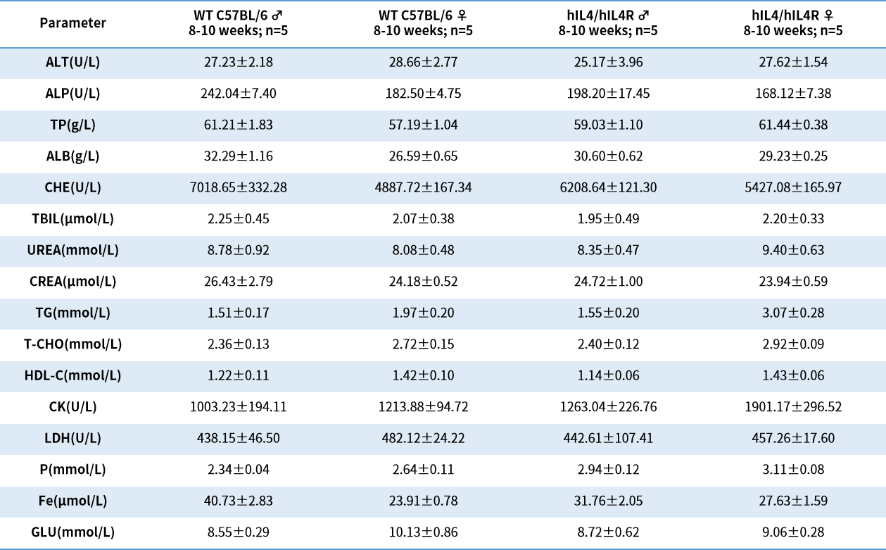
Abbr. WT, wild type; HO, homozygous.
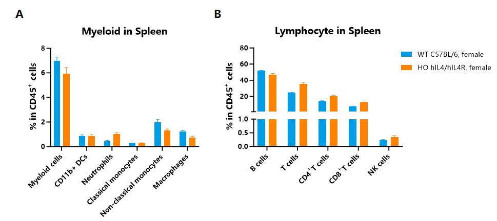
There were no obvious pathological changes in these tissues (n=3, 8-10 weeks old, 100x magnification).
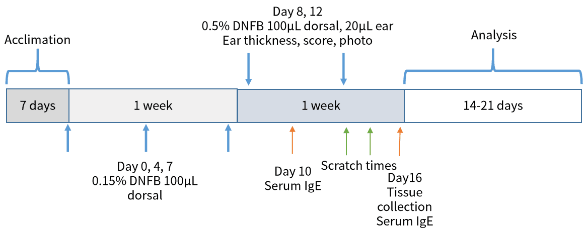
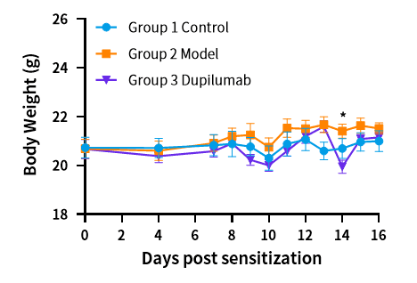
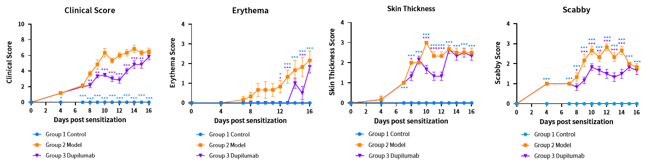
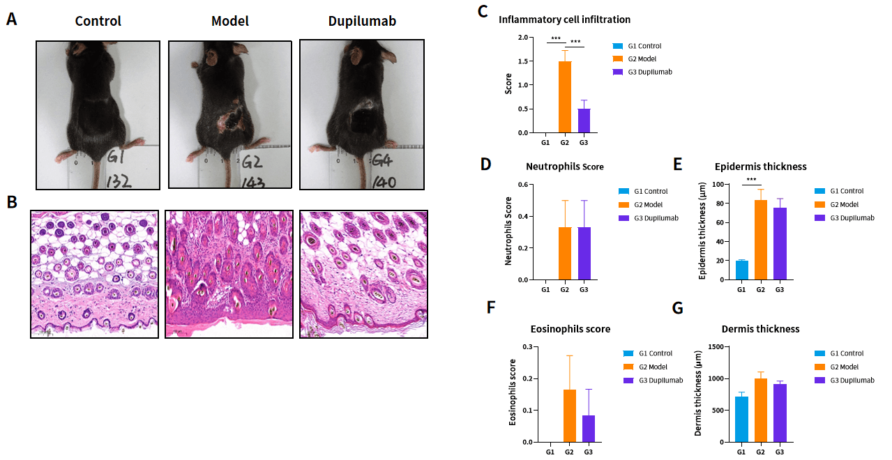
Case 1: In vivo efficacy of anti human IL4RA mAb in the DNFB induced Atopic dermatitis Model based on hIL4/hIL4R Mice

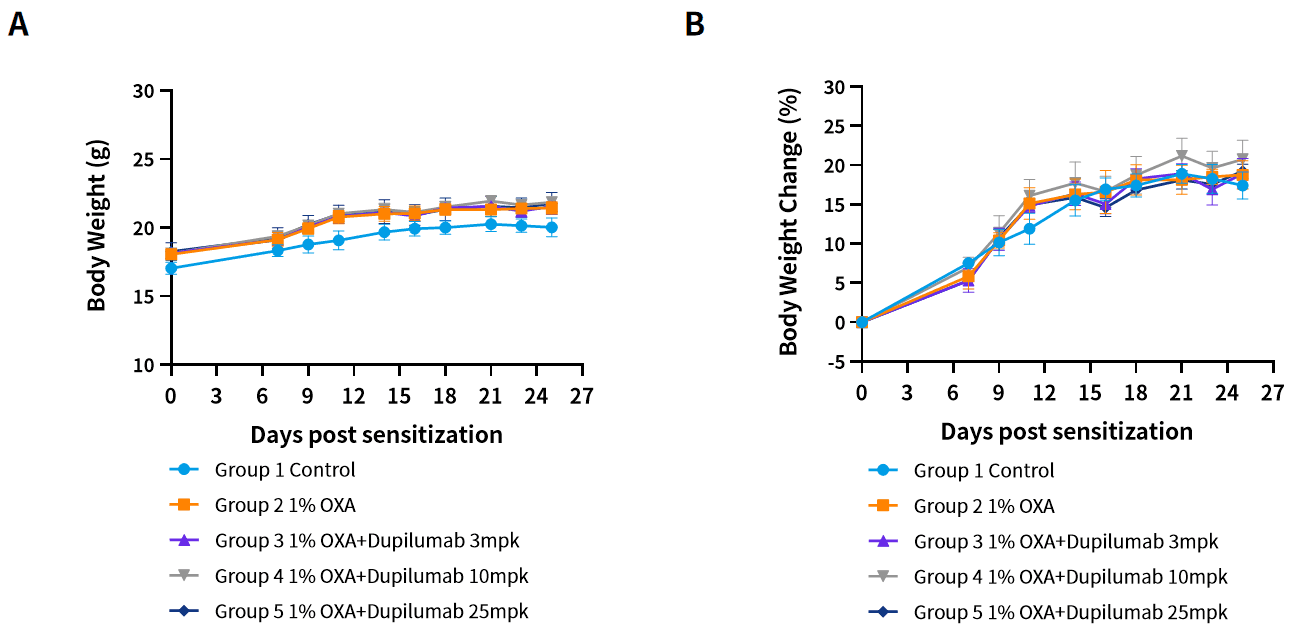
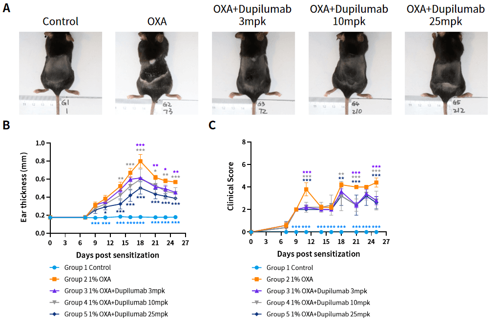
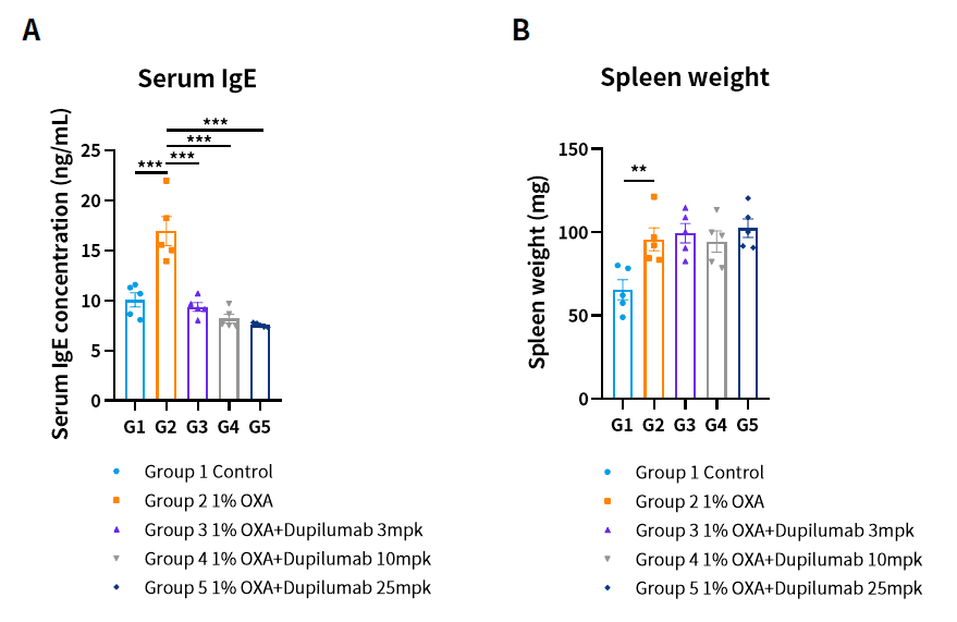
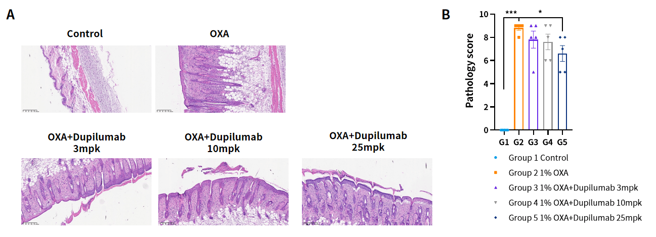
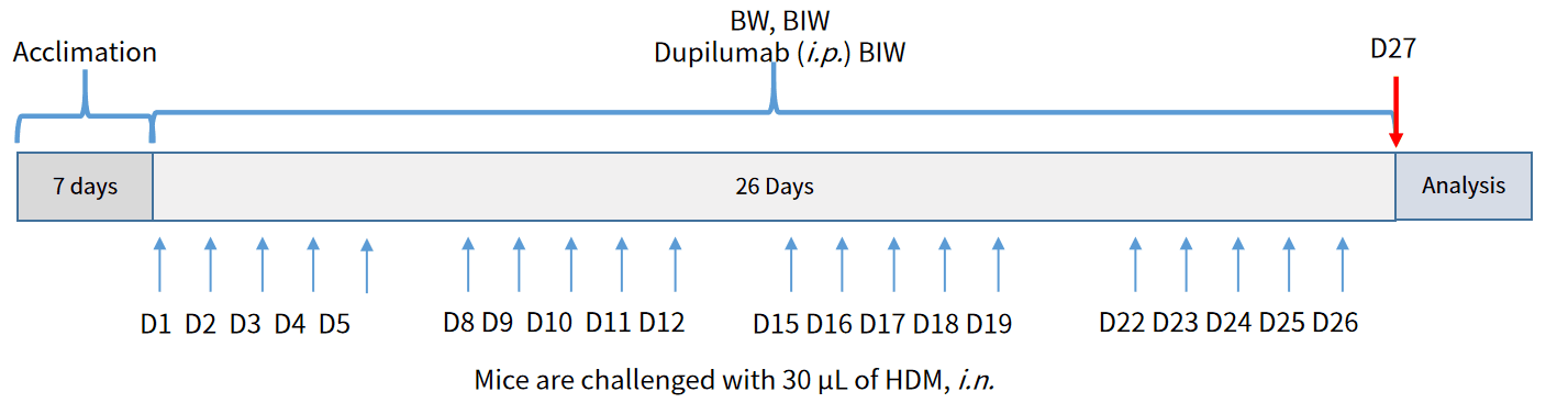
Case 2: In vivo efficacy of anti human IL4RA mAb in the OXA induced Atopic dermatitis Model based on hIL4/hIL4R Mice
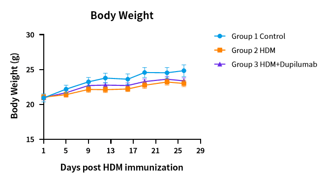
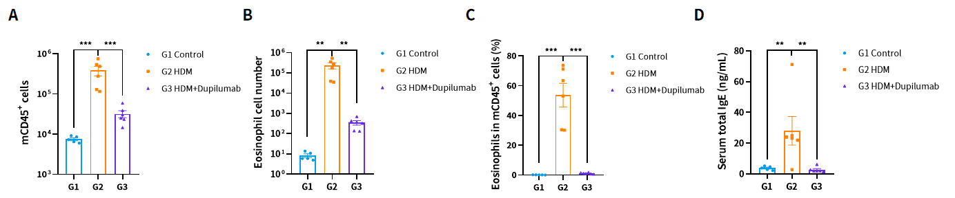
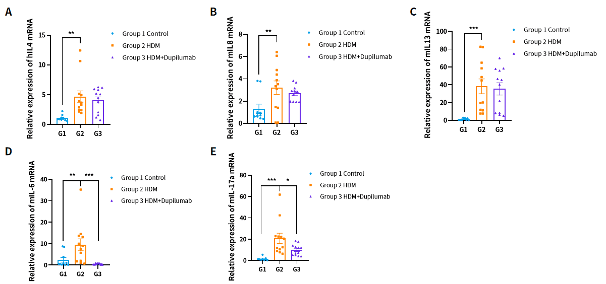
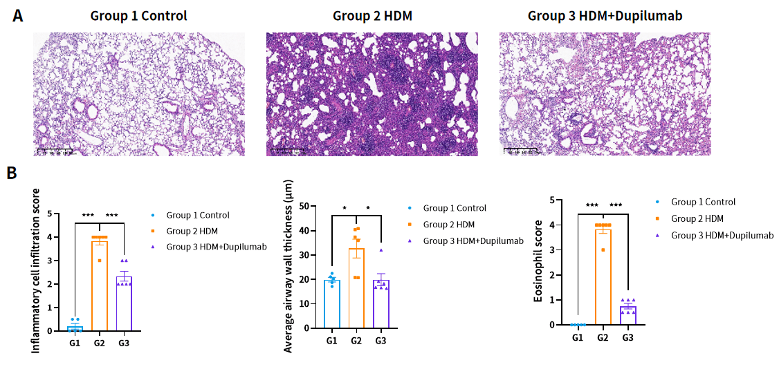
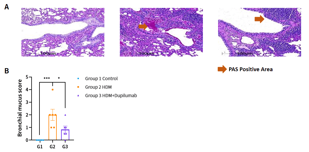
Case 3: In vivo efficacy of anti human IL4RA mAb in the HDM induced Asthma Model based on hIL4/hIL4R Mice
About ingenious targeting laboratory
Ingenious targeting laboratory maintains a catalog of over 14,774 mouse models, including humanized strains, Cre driver lines for conditional expression, and reporter mice for cell tracking and imaging. These quality-controlled models on defined genetic backgrounds ship as breeding pairs or cohorts with complete genotyping protocols and health documentation. Researchers gain immediate access to mouse strains without custom generation timelines, accelerating experiments across immunology, oncology, neurology, and metabolic disease applications. If you are interested in our hIL4/hIL4R mouse model, please contact us. Or, please search our catalog for your gene of interest.